Two research projects have recently benefited from RCC eResearch Analyst Dr Nick Hamilton’s weekly Image Clinic at UQ.
Nick helped PhD student Vikram Narayan, who works in Deputy Associate Dean of Science Professor Steve Chenoweth’s lab, automate a workflow that would have previously taken hours and hours of manual labour.
The Chenoweth Lab, which aims to test fundamental hypotheses in genetics and evolutionary biology, is studying the genomes of fruit flies, known scientifically as Drosophila serrata.
These flies have two distinct genotypes, distinguished by the flies having either red or orange eyes, and the researchers need to count how many of each there are.
Vikram is measuring the lifetime reproductive success of individual flies by distinguishing between red and orange-eyed offspring.
“These differences in fitness help us to understand the roles of natural selection and adaptation on life history traits such as lifespan,” said Vikram.
“However, for species with multiple reproductive cycles or large numbers of offspring, measuring lifetime reproductive success is often arduous and time-consuming, resolved only by the most labour-intensive studies.”
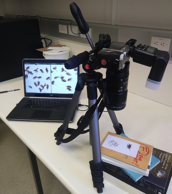
“Currently, the researchers are manually counting thousands of flies, and plan to do tens of thousands more in the future which is painstaking work,” said Nick. “So, I created a hardware camera setup for imaging the flies at high resolution, and then an analytic pipeline to find and count the eyes.”
“With Dr Nick’s help, I now hope to be able to do in days or weeks, what would previously have taken months and months of intense labour,'' said Vikram.
Nick has also helped early-career researchers Ryan Vandenberg Brady and Tish Essebier who work in Dr Emma Gordon's Vessel Dynamics Lab at the Institute for Molecular Bioscience (IMB).
Ryan and Tish wanted to distinguish changes and the seven classes of cell boundaries when the cells are treated by different molecules.
Nick created a pipeline that “chopped up” the cell images into small regions and then automatically classified each of the regions using machine deep learning methods.
He used RCC-managed high-performance computer Wiener to train the machine learning model for the cell imaging. The trained model now runs on one of the Vessel Dynamics Lab’s laptops.
"Dr Nick's model helped us to understand a key pathway that controls blood vessel formation. We were able to identify changes in how cells attach to each other when a key signalling molecule was mutated. The model continues to bring us closer to understanding diseases such as retinopathy of prematurity and diabetic retinopathy," said Tish.
The Vessel Dynamics Lab’s research is focused on the formation and maintenance of the blood and lymphatic vascular systems.
The lab uses novel biological models, biochemical assays and imaging techniques to better understand vessel biology, which will enable improved treatment of disease and aid in the development of vascularised, bioengineered organs.
Dr Nick’s Image Clinic is held most Monday mornings at IMB. See the RCC website for more information.


